Fused, real-time, gamma-optical overlay
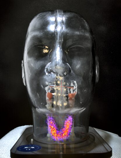
Seracam® forms images that reveal the distribution of a radiopharmaceutical in various organs to help diagnose and monitor disease. Seracam® uses a microcolumnar CsI(Tl) crystal scintillator to convert gamma photons to optical photons that are detected by a semi-conductor.
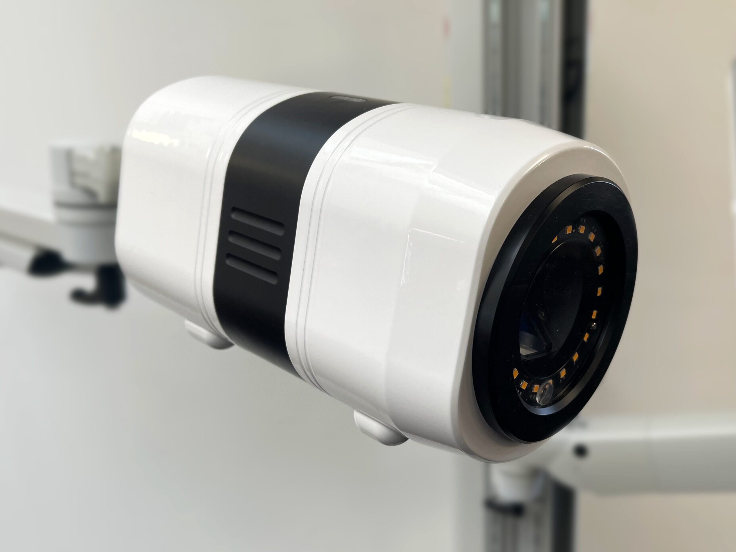
Incoming gamma rays are collimated by a pinhole collimator in order to preserve image resolution. Seracam includes four different pinholes that can be automatically selected to optimise the balance of image resolution and speed of image acquisition for the case at hand – a novel feature unique to Seracam.
An optical camera is built-in with the same field of view as the gamma detector, overlaid and displayed on the control PC in real time with alignment maintained at any angle or imaging distance, another industry first.
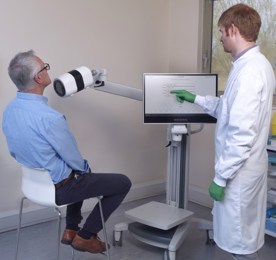
This technology is anticipated to make images easier to interpret for the untrained eye, which should enhance physician patient communication and help patients gain a better understanding of their conditions and treatment options.
Performance testing results demonstrate that the gamma-imaging performance of Seracam, in aspects such as spatial resolution, spatial linearity and uniformity values, exceeds that of large field-of-view cameras. For more details see here.